Jonatan Merino-Soriano1 ID· José Luis Gálvez Romero2 ID· Andrea Báez Núñez3 ID· Sayuri Chong Hernández4 ID· Francisco de Jesús García-Mendoza5 ID
1Department of Internal Medicine. Hospital Regional de Puebla, Instituto de Seguridad y
Servicios Sociales de los Trabajadores del Estado.
2Department of Research. Hospital
Regional de Puebla, Instituto de Seguridad y Servicios Sociales de los Trabajadores del
Estado.
3Department of Nephrology. Hospital Regional de Puebla, Instituto de
Seguridad y Servicios Sociales de los Trabajadores del Estado.
4Department of
Internal Medicine. Hospital Regional de Puebla, Instituto de Seguridad y Servicios Sociales de los
Trabajadores del Estado.
5Central Research Laboratory. Faculty of Medicine,
Benemérita Universidad Autónoma de Puebla.
Puebla, México.
Acta Gastroenterol Latinoam 2026;56(3):304
Received: 25/06/2026 / Accepted: 31/08/2026 / Published online: 30/09/2026 / https://doi.org/10.52787/agl.v56i3.667
A 66-year-old woman with a history of type 2 diabetes mellitus, hypertension, and advanced chronic kidney disease on peritoneal dialysis was admitted with peritoneal dialysis-associated peritonitis. During hospitalization, she developed intolerance to oral intake, progressive dysphagia, epigastric pain, and coffee-ground emesis. Laboratory evaluation revealed moderate normocytic normochromic anemia, leukocytosis with neutrophilia, hypoalbuminemia (2.1 g/dL), and hyperglycemia (up to 303 mg/dL). Peritoneal fluid analysis showed 1,130 leukocytes/mm³ with 78% polymorphonuclear cells and Gram-negative bacilli on Gram staining, supporting the diagnosis of peritoneal dialysis-associated peritonitis.
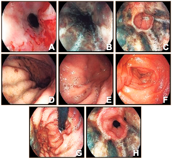
Given the suspicion of upper gastrointestinal bleeding, upper gastrointestinal endoscopy was performed on November 22, 2024, revealing extensive circumferential involvement of the distal esophageal mucosa with a sharply demarcated transition at the gastroesophageal junction (Figure 1). Based on the clinical context and endoscopic findings, what is the most likely diagnosis?
Figure 1.
Solución del caso en la página 365
Correspondence: Jonatan Merino-Soriano
Email: jmerino96@hotmail.com
Acta Gastroenterol Latinoam 2026;56(3):304