performed on September 23, 2025
The image corresponds to acute esophageal necrosis, also known as “black esophagus.” As illustrated in Figure 1B, upper gastrointestinal endoscopy demonstrated diffuse circumferential black discoloration of the distal esophageal mucosa with a well-demarcated transition at the gastroesophageal junction, a hallmark feature of acute esophageal necrosis. This condition is usually associated with severe underlying illnesses and the combination of multiple predisposing factors that contribute to esophageal mucosal injury. In this patient, sepsis secondary to peritonitis, advanced chronic kidney disease on peritoneal dialysis, type 2 diabetes mellitus, anemia, and hypoalbuminemia likely contributed to its development. Conservative treatment was initiated, including bowel rest, proton pump inhibitors, sucralfate, and management of the underlying infection, resulting in progressive clinical improvement. Early recognition of this condition is essential to reduce the risk of complications and guide timely treatment.
The patient showed progressive clinical improvement during hospitalization after initiation of conservative management. Coffee-ground emesis, dysphagia, and epigastric pain gradually resolved, and oral intake was progressively reintroduced. A liquid diet was initially tolerated and subsequently advanced as symptoms improved. No clinical evidence of early complications of acute esophageal necrosis, including esophageal perforation or mediastinitis, was identified.
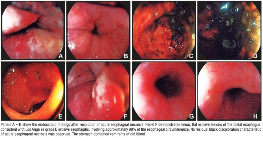
Given the sustained clinical improvement, resolution of upper gastrointestinal bleeding symptoms, progressive tolerance of oral intake, and absence of signs suggestive of esophageal perforation or other complications, repeat upper gastrointestinal endoscopy was not performed during the same hospitalization. The patient was discharged with outpatient follow-up by the Gastroenterology and Nephrology departments. During outpatient follow-up, an upper gastrointestinal endoscopy requested by the Gastroenterology Department was performed on September 23, 2025. The examination showed no residual endoscopic features of acute esophageal necrosis. Instead, severe Los Angeles grade D erosive esophagitis was identified, with linear, flat erosions greater than 5 mm involving approximately 90% of the esophageal circumference and along with remnants of old blood in the stomach (Figure 2). Proton pump inhibitor therapy was therefore continued.
During a subsequent hospitalization for upper gastrointestinal bleeding, imaging studies demonstrated findings consistent with portal hypertension. Upper gastrointestinal endoscopy performed on October 27, 2025, revealed Los Angeles grade C erosive esophagitis and chronic gastropathy, without evidence of recurrent acute esophageal necrosis (Figure 3). The patient showed a favorable clinical response to medical management and was discharged on pharmacological therapy with continued outpatient follow-up.
Figure 2. During outpatient follow-up, a control upper gastrointestinal endoscopy requested
by the Gastroenterology Department was
performed on September 23, 2025
Figure 3. Upper gastrointestinal endoscopy performed on October 27, 2025, during a subsequent
hospitalization for upper gastroin-
testinal bleeding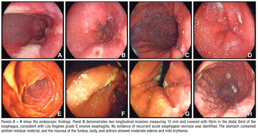
Consent for publication. The authors certify that informed consent was obtained from the patient for the publication of her clinical information and images. The patient understands that her name and initials will not be published and that all reasonable efforts will be made to protect her identity; however, complete anonymity cannot be guaranteed. Approval by an Institutional Review Board or Research Ethics Committee was not required for the publication of this case report.
Intellectual property. The authors declare that the data and figures presented in this manuscript are original and were carried out at their institutions.
Funding. The authors declare that there were no external sources of funding.
Conflict of interest. The authors declare that they have no conflicts of interest in relation to this article.
Copyright

© 2026 Acta Gastroenterológica latinoamericana. This is an open-access article released under the terms of the Creative Commons Attribution (CC BY-NC-SA 4.0) license, which allows non-commercial use, distribution, and reproduction, provided the original author and source are acknowledged.
Cite this article as: Merino-Soriano J, Gálvez Romero J L, Báez Núñez A et al. An Unusual Distal Esophageal Finding: An Endoscopic Challenge. Acta Gastroenterol Latinoam. 2026;56(3):365. https://doi.org/10.52787/agl.v56i3.667
Acta Gastroenterol Latinoam 2026;56(3):365