
Evaluation of a Patient with Epigastric Pain and Uncommon Synchronous Findings
DOI:
https://doi.org/10.52787/agl.v55i4.558Keywords:
Epigastric pain, uncommon synchronous findings, tomographyAbstract
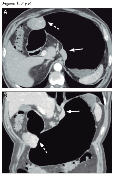
A 64-year-old male patient, with no relevant medical history, presented with subacute epigastric pain. Laboratory tests revealed decreased eosinophils and lymphocytes, with mild thrombocytopenia as the only abnormal finding. Due to dyspeptic symptoms, an upper gastrointestinal endoscopy was performed, which showed two gastric lesions: one in the subcardial region, with regular borders and intact mucosa, and a larger lesion in the distal gastric body, also with preserved mucosa. Based on the endoscopic findings, an abdominal CT with CO2 gastric distension (pneumotomography), including three-dimensional reconstructions, was requested. Additionally, a virtual endoscopy generated through post-processing was performed, allowing for a more precise characterization of the lesions’ morphology, exact location, and growth pattern.
References
-1. Kang HC, Menias CO, Gaballah AH, Shroff S, Taggart MW, Garg N, et al. Beyond the GIST: mesenchymal tumors of the stomach. Radiographics. 2013 Oct;33(6):1673-90.
-2. Lin YM, Chiu NC, Li AFY, Liu CA, Chou YH, Chiou YY. Unusual gastric tumors and tumor-like lesions: Radiological with pathological correlation and literature review. World J Gastroenterol. 2017 Apr 14;23(14):2493-504.
Downloads
Published
How to Cite
Issue
Section
License
Copyright (c) 2025 Roy López Grove, Daniela Soloaga, Juan Carlos Spina

This work is licensed under a Creative Commons Attribution-NonCommercial-ShareAlike 4.0 International License.


